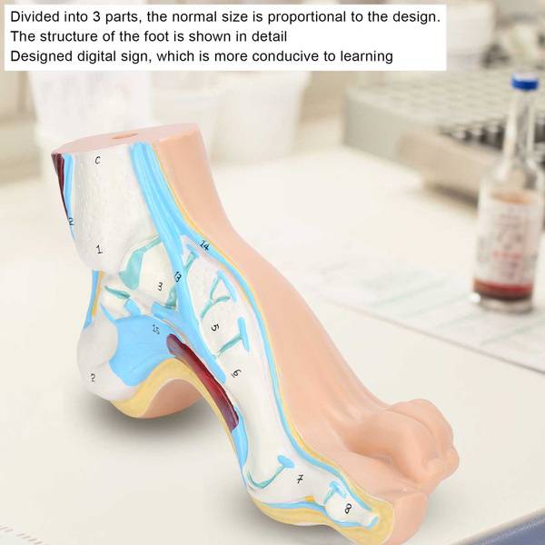
Human Foot Model
$517
<section>
Option 1: Anatomical Human Foot Model with Arch Variations
Description: Discover the intricacies of the human foot with this highly detailed anatomical model by Xinyanr. It meticulously illustrates the bone, muscle, and ligament structures of a normal foot, flatfoot, and high-arched foot, providing invaluable insights for learning and demonstration. Featuring an innovative digital labeling system for enhanced understanding, this 1:1 adult-sized model is crafted from durable, eco-friendly PVC. Its versatility makes it ideal for medical education, lectures, presentations, and research, offering a natural display with superficial fascia removed to reveal underlying musculature and ligaments. This rare, practical teaching tool facilitates a deeper comprehension of foot anatomy and biomechanics, crucial for surgical medicine and foot research.
</section> <hr> <section>Option 2: Realistic 3D Foot Anatomy Model for Medical Training
Description: Elevate your anatomical studies with the Xinyanr Human Foot Model, a premium 1:1 adult-sized educational tool. This durable, eco-friendly PVC model showcases the skeletal muscles and ligaments of normal, flat, and high arches with exceptional clarity. Its three-part design and convenient digital marking system streamline learning for students, educators, and medical professionals. Perfect for lectures, presentations, and in-depth foot research, this model accurately depicts the natural form of the foot, with superficial fascia removed to expose critical structures for a comprehensive understanding. A vital resource for surgical medicine and biomechanical analysis of the foot.
</section> <hr> <section>Option 3: 3-Part Human Foot Anatomy Model: Normal, Flat, High Arch
Description: Explore human foot anatomy with this comprehensive 1:1 adult-sized model. Xinyanr's eco-friendly PVC model clearly displays the bones, muscles, and ligaments of normal, flat, and high-arched feet. Its practical 3-part design and integrated digital labeling system enhance educational effectiveness. Ideal for medical training, lectures, and presentations, it offers a realistic, detailed view with superficial fascia removed, revealing key anatomical structures for research and learning. A superior tool for understanding foot biomechanics and surgical considerations.
</section>